Educational resource developed at the Veterinary Anatomy Section, ICAR-Indian Veterinary Research Institute (ICAR-IVRI), Bareilly.
📝 STANDARD DESCRIPTION:
Avian Wing (Fowl): - Pectoral girdle = Scapula + Coracoid + Clavicle. - Clavicles fuse ventrally to form the Furcula (wishbone). - Humerus is pneumatic (air-filled). - Distal carpals + metacarpals fuse into a single Carpometacarpus.
📚 DETAILED DESCRIPTION:
FORELIMB OF THE FOWL — THE WING
The wing keeps the same basic segments as the mammalian forelimb (girdle, brachium, antebrachium, manus), but the bones of the girdle and the manus are reduced in number and fused for rigidity.
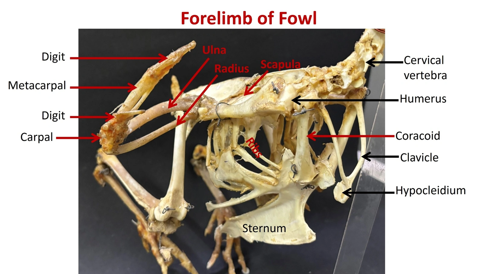
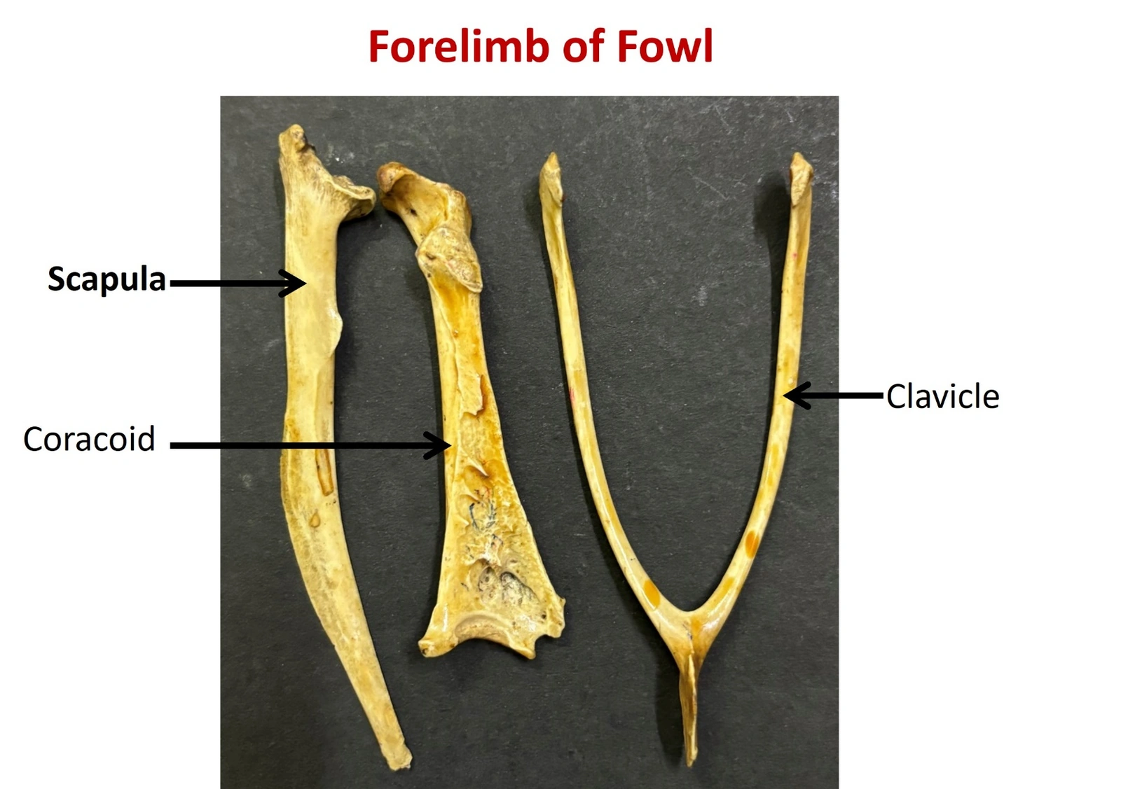
PECTORAL (SHOULDER) GIRDLE — THREE BONES: • Scapula: long, narrow and sabre-like; lies almost horizontally along the dorsal thorax, parallel to the vertebral column. • Coracoid: short, stout and the strongest bone of the girdle; forms a rigid strut from the shoulder to the sternum. • Clavicles: the two clavicles fuse at the midline to form the furcula ('wishbone'). Its ventral median projection is the hypocleidium.
TRIOSSEAL CANAL: Where the scapula, coracoid and clavicle meet, a foramen called the triosseal canal is formed. The tendon of the supracoracoideus muscle passes through it and turns over the shoulder to insert on the dorsal humerus, so this ventral muscle raises (elevates) the wing.
SHOULDER JOINT: The glenoid cavity is formed jointly by the scapula and the coracoid.
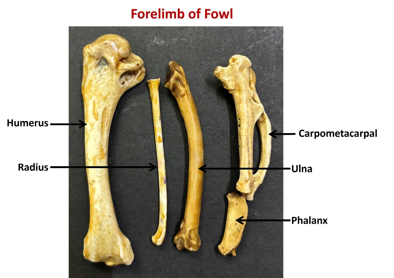
BRACHIUM — HUMERUS: • A pneumatic bone: a pneumatic foramen in the pneumatic fossa on the proximal caudal aspect admits a diverticulum of the clavicular air sac. • Proximal end carries the head and prominent deltoid and ventral tubercles for the powerful flight muscles.
ANTEBRACHIUM — RADIUS & ULNA: • Radius: the slender, straighter of the two, lying cranially. • Ulna: stouter and bowed (caudal). Its caudal border bears small papillae (quill knobs) for the secondary flight feathers. • The two bones remain separate, allowing the wing to fold and extend.
MANUS — CARPUS, CARPOMETACARPUS AND DIGITS: • Only two free carpal bones remain — the radiale and the ulnare. • The distal carpals fuse with the metacarpals to form the single carpometacarpus. • Three reduced digits are present: the alular digit (carries the alula or 'bastard wing'), the major digit and the minor digit.
STERNUM (associated): The sternum bears a deep median keel (carina) that greatly enlarges the area of attachment for the pectoralis and supracoracoideus muscles.
Girdle is reduced to the scapula alone (clavicle absent or vestigial); the limb is slung to the trunk by muscle. No coracoid strut and no furcula.
FOWL
Girdle has three bones. The coracoid braces the shoulder against the sternum and the fused clavicles (furcula) act as a spring between the shoulders during wing beats.
FOWL
Carpus has only two free bones (radiale, ulnare) and the manus is fused into a carpometacarpus, whereas the ox has six and the horse seven or eight carpal bones.
CLINICAL RELEVANCE
Intraosseous fluid therapy: Use the distal ulna or the proximal tibiotarsus. Never use the humerus or femur — they are pneumatic and communicate with the air sacs, so injected fluid would enter the respiratory system. Wing fractures: The radius and ulna are commonly fractured; because the ulna carries the secondary feathers, callus or malunion here affects flight. Furcula and keel: The keel is used for body-condition scoring in poultry; a prominent keel indicates loss of pectoral muscle mass.
VISUAL REFERENCE
Fowl
Articulated shoulder girdleForelimb of Fowl — Shoulder girdle in situShoulder girdle bonesForelimb of Fowl — Scapula, coracoid and clavicleWing bonesForelimb of Fowl — Humerus, radius, ulna, carpometacarpus, phalanx
BIOMECHANICS ENGINE
Comparative Veterinary Biomechanics
The Why of Veterinary Anatomy
Explore the hidden mechanics of why structures are present, absent or modified across domestic and wildlife species—and how each anatomical design supports movement, function and clinical practice.
HIGH-QUALITY VETERINARY IMAGES IN DEVELOPMENTCarefully curated visuals to support anatomical learning will be available by September 2026.
Category
Structure Name
Vs. Tag
The Biomechanical "Why"
Explanation goes here.
Clinical Relevance
Clinical notes go here.
ENGINEERING CHALLENGE
Choose your arena
How many questions?
Q1/10
Score0
Streak0
Topic
Loading challenge...
Session Complete
8 / 10
Excellent
Best streak0
Accuracy0%
TopicAll
SELECT REGIONAL ANATOMY
STEP 1 OF 3
SELECT SYSTEM
REGION > SYSTEM
SELECT CHALLENGE FORMAT
REGION > SYSTEM > FORMAT
MULTIPLE CHOICE
0 questions
Select the best answer from options
TRUE / FALSE
0 questions
Test your knowledge with binary choices
FILL IN BLANKS
0 questions
Type the correct answer
EXAM MODE
Timed mock exam
Customisable: pick duration + question count + format. No instant feedback — like a real exam.
Configure Your Exam
REGION > SYSTEM
Available: ? questions in this pool
Q 1/10
RegionSystem
00:00
Loading...
PrevNext
Question Navigator
Current Correct Incorrect Unanswered Bookmarked
PERFORMANCE ANALYSIS
SESSION REPORT
0%
ACCURACY
0
CORRECT
0
INCORRECT
0
UNANSWERED
0m 0s
TIME TAKEN
PROFESSIONAL RECOMMENDATION:
...
Category Breakdown
DETAILED REVIEW
PERFORMANCE DASHBOARD
Accuracy Trend
Last 20 sessions
Topic Strength Map
Accuracy by Syllabus Unit × Topic
Recent Quiz History
Your last sessions
Welcome back, student
Your study companion for B.V.Sc Anatomy
0Bookmarks
0Highlights
0Notes
0Read
0
day streak
0longest
0total days
Last 12 weeks
LessMore
Navigation position Choose where the menu bar sits on desktop. Mobile always stays at the bottom.
Why this matters: Your bookmarks, notes, highlights, and quiz progress live in your browser only.
If you clear browser data, switch phones, or use incognito by accident, they're gone forever.
Download a backup at least once a month and keep the JSON file in Google Drive / WhatsApp to yourself.
Veterinary Anatomy Studio
EXPLORING ANATOMY THROUGH TECHNOLOGY.
Overview
An interactive, curriculum aligned veterinary anatomy platform for B.V.Sc., M.V.Sc. and DVM students worldwide. Developed at ICAR–Indian Veterinary Research Institute, Bareilly, it brings together regional anatomy, histology, embryology, comparative biomechanics, clinical relevance, quizzes and revision.
Vision
To make veterinary anatomy approachable, comparative, and clinically grounded — for every B.V.Sc student, on every device, even offline.
Mission
Cover the complete VCI Unit 1–8 syllabus with high-resolution comparative content.
Pair every structure with its biomechanical "why" and clinical correlation.
Make active recall the default — MCQ, True/False, Fill-in, Smart Review, Exam Mode.
Stay free, ad-free, and lightweight enough to run on any phone.